In short
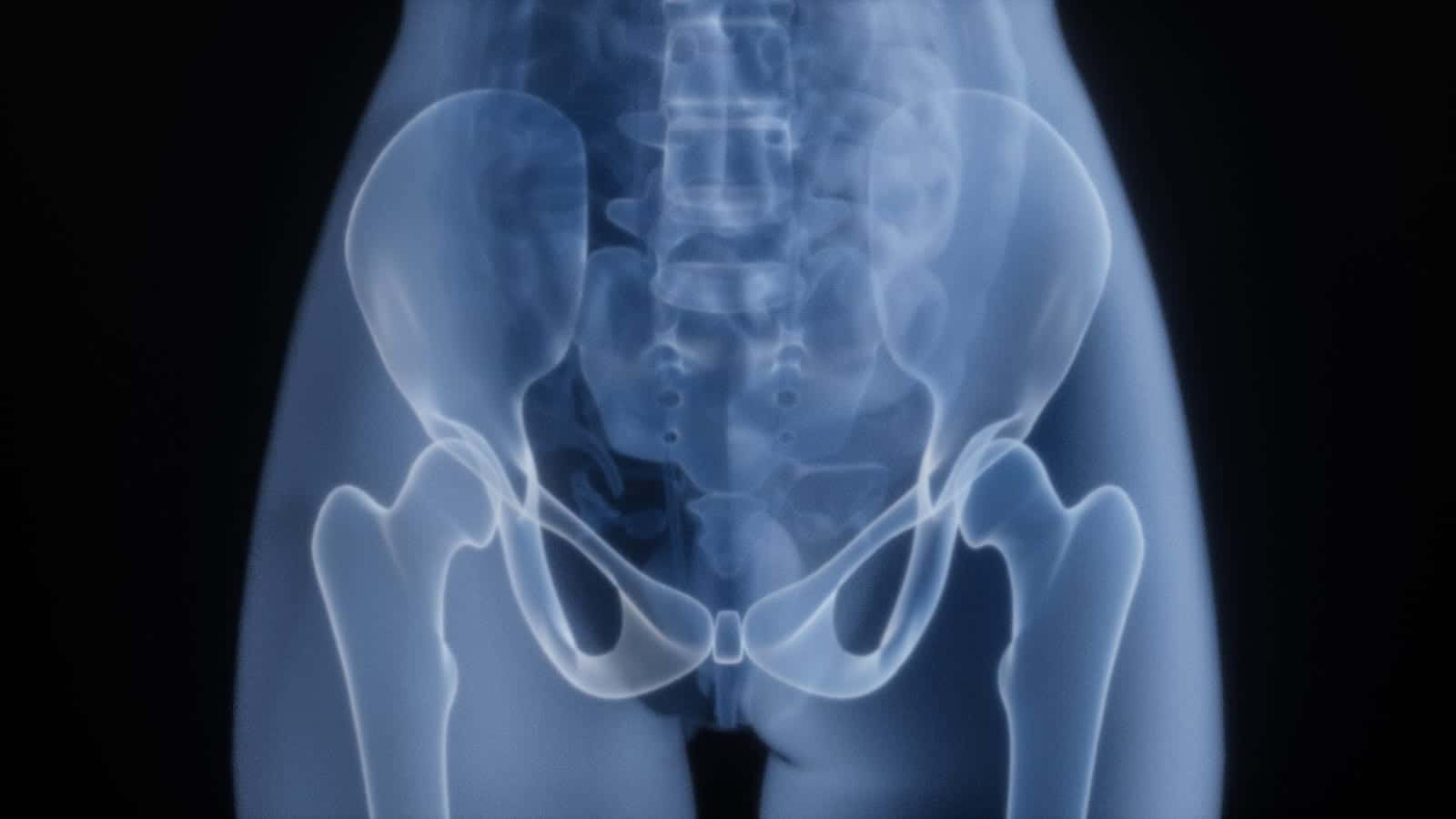
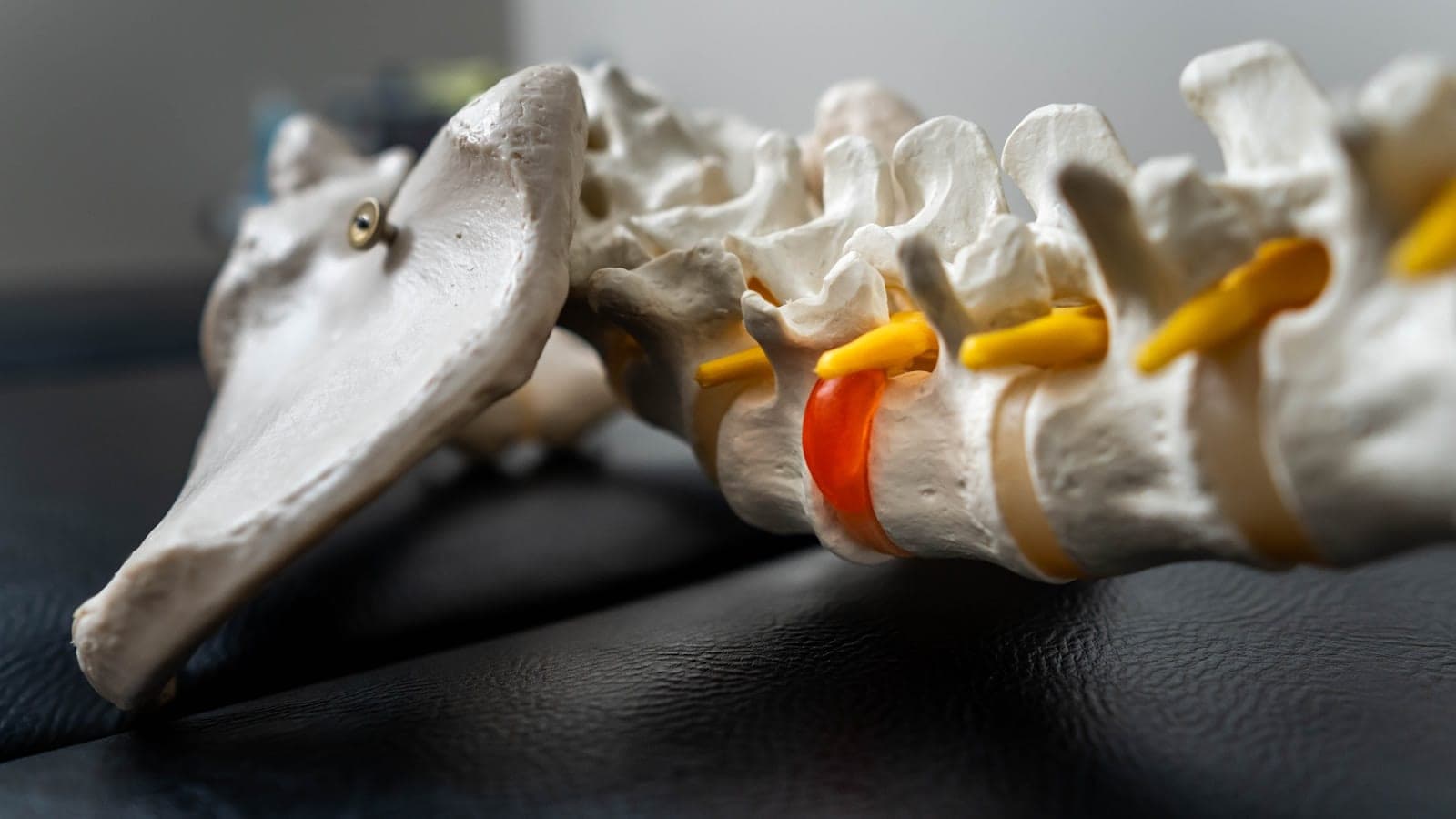
Ankylosing spondylitis is a chronic inflammatory arthritis that targets the sacroiliac joints and spine. Inflammation at the entheses — where ligaments and tendons anchor into bone — is followed by new bone formation, which can eventually fuse spinal segments.
- Category
- Spondyloarthritis
- Seen first as
- Low back and buttock pain before age 40, developing gradually over months
- Treated with
- Daily structured exercise, the foundation
What is Ankylosing Spondylitis?
It typically begins in the late teens to early thirties, and it is the classic cause of back pain in a young adult that gets dismissed as a gym injury or a bad mattress for five to ten years.
The distinguishing feature is inflammatory back pain: gradual onset, worse in the second half of the night, more than 30 minutes of morning stiffness, better with movement and no better with rest.
 Illustrative
In the Indian context
COPCORD data put spondyloarthritis prevalence in India at roughly 0.23%. HLA-B27 is common in the general Indian population, so testing positive without inflammatory symptoms is not a diagnosis, a point of frequent and expensive confusion.
Symptoms of Ankylosing Spondylitis
Early signs
- Low back and buttock pain before age 40, developing gradually over months
- Pain and stiffness worst on waking or after prolonged sitting; better after a hot shower or activity
- Alternating buttock pain that shifts from side to side
- Heel pain at the Achilles insertion or under the sole (enthesitis)
- A swollen finger or toe that looks like a sausage (dactylitis)
If left untreated
- Reduced spinal flexion, chest expansion below 5 cm, and stooped posture
- Neck stiffness and difficulty looking over the shoulder while driving
- Recurrent painful red eye with light sensitivity (acute anterior uveitis)
- Associated inflammatory bowel disease or psoriasis
- Osteoporotic spine fracture from relatively minor trauma
Causes & risk factors
- HLA-B27 positivity — present in 80–90% of patients, though most B27-positive people never develop the disease
- Family history of AS, psoriasis, uveitis or inflammatory bowel disease
- Gut mucosal inflammation and microbiome changes driving joint inflammation
- IL-17 and IL-23 immune pathway activation
- Male sex — men tend to develop more radiographic fusion, though women are affected as often and are diagnosed later
How Ankylosing Spondylitis is diagnosed
No single test diagnoses a rheumatic disease. The diagnosis comes from combining the pattern of symptoms, the examination findings, and only the investigations that answer a specific question.
 Illustrative
Inflammatory back pain assessment
A structured history using ASAS criteria — age of onset, night pain, morning stiffness duration, response to exercise versus rest — carries more diagnostic weight than any blood test.
MRI of the sacroiliac joints
STIR sequences show bone marrow oedema years before X-ray changes appear. This is what allows non-radiographic axial spondyloarthritis to be diagnosed and treated early.
X-ray sacroiliac joints and spine
Detects established sacroiliitis, squaring of vertebrae and syndesmophytes. Normal X-rays do not exclude early disease.
HLA-B27 typing
Interpreted strictly in context. It raises probability substantially in a patient with inflammatory back pain, and means little in a patient with mechanical pain.
CRP, ESR and BASDAI scoring
Objective baselines. BASDAI and ASDAS scores are what guidelines and insurers use to justify escalation to biologics.
Screening for extra-articular disease
Eye, skin and bowel review at every visit, since uveitis, psoriasis and IBD change the choice of drug.
Treatment options
 Illustrative
Daily structured exercise, the foundation
Spinal extension, rotation and deep-breathing exercises done consistently are as important as medication. Physiotherapy-led programmes measurably preserve posture and chest expansion.
NSAIDs taken continuously, not on demand
In AS, regular full-dose NSAIDs are both symptomatic and possibly structure-modifying. Gastric and renal protection is planned alongside.
Biologic therapy
TNF inhibitors and IL-17 inhibitors such as secukinumab and ixekizumab transform disease that fails two NSAIDs. Choice is guided by uveitis or IBD history.
JAK inhibitors
Oral upadacitinib and tofacitinib are approved options for active axial disease.
Targeted enthesitis and peripheral joint care
Local injections for persistent enthesitis; sulfasalazine or methotrexate where peripheral joints dominate. Neither helps axial disease.
Bone health and posture protection
AS paradoxically causes osteoporosis. DEXA scanning, vitamin D, calcium and fall precautions matter, because a fused spine fractures easily.
Medicine guides
How to take them, side effects, blood tests and pregnancy, for the medicines most often used for this condition.
Living with Ankylosing Spondylitis
- Sleep flat on a firm mattress with a thin pillow; avoid curling into a foetal position
- Set the monitor at eye level — office posture accelerates cervical stiffening
- Swimming is the single best exercise for AS: extension, rotation and cardio without impact
- Stop smoking. It is strongly linked to faster spinal fusion and worse function
- Practise deep breathing daily to maintain chest wall expansion
Seek urgent medical attention if you have
- Sudden new severe neck or back pain after even minor trauma, a fused spine fractures unstably
- A red, painful, light-sensitive eye — see an ophthalmologist the same day
- New leg weakness, numbness, or bladder or bowel changes
- Persistent fever and weight loss — spinal infection or TB must be excluded, especially in India
- Chronic diarrhoea or blood in stool suggesting inflammatory bowel disease
Do not wait for a scheduled appointment for any of the above. Call the clinic on +91 70571 19999 or attend the nearest emergency department.
Frequently asked questions about Ankylosing Spondylitis
I am HLA-B27 positive. Will I get ankylosing spondylitis?
Will my spine definitely fuse?
Why does my back hurt more when I rest?
Can I go to the gym?
Get an expert opinion on Ankylosing Spondylitis in Pune
Consultations at the Ravet clinic in Pimpri-Chinchwad. Bring your previous reports — a properly reviewed history usually saves repeating tests.
Part of a wider group
Spondyloarthritis & Back PainBack pain that is worse at rest and better with movement is not a mechanical strain.How ankylosing spondylitis is treated at the clinic
Ankylosing Spondylitis is managed under spondyloarthritis & back pain — the same pathway, targets and monitoring schedule described in full on that page.
Related conditions
Psoriatic Arthritis
When psoriasis moves into the joints, often years after the first skin patch.
Reactive Arthritis
Arthritis that follows an infection elsewhere, usually self-limiting, occasionally not.
Osteoporosis
Silent until the first fracture, which is exactly why it should be screened for, not waited for.
Rheumatoid Arthritis
Symmetric small-joint swelling that responds best when treated within the first 12 weeks.